
Intro to Knee Ultrasound
Knee ultrasound can be used to visualize important anatomical structures and to visualize possible tendon or muscular injuries.
On ultrasound, it is important to know that bone is hyper-echoic, or bright white. Ultrasound waves cannot penetrate bone, and instead reflect off the bony cortex, so bone appears as a bright white line, and any structures deep to the cortex of bone will be dark.
Now let's start with the anatomy.
In a knee ultrasound some of the relevant structures we may see include:
-
Bones
-
Femur
-
Patella
-
Tibia
-
Fibula
-
-
Tendons
-
Patellar
-
Quadriceps
-
-
Ligaments
-
Medial Collateral Ligament
-
Lateral Collateral Ligament
-
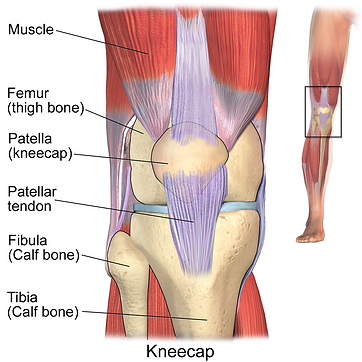
Blausen.com staff (2014). "Medical gallery of Blausen Medical 2014". WikiJournal of Medicine 1 (2). DOI:10.15347/wjm/2014.010. ISSN 2002-4436., CC BY 3.0, via Wikimedia Commons
How to get the Sagittal Views
(Suprapatellar or Infrapatellar):
-
It is easiest to have the patient in a supine position, with their knee slightly flexed
-
Place the probe in a sagittal or longitudinal plane with the indicator pointing superiorly, toward the hip
-
Start with the probe on the lower thigh, above the knee joint, and scan inferiorly toward the knee joint until you have scanned through the entire joint, and are now at the location of the superior tibia


How to get the Axial Views
(Suprapatellar or Infrapatellar):
-
Position the patient as above
-
Place the probe in an axial or transverse plane with the indicator pointing laterally
-
Start with the probe on the lower thigh, above the knee joint, and scan inferiorly toward the knee joint until you have scanned through the entire joint, and are now at the location of the superior tibia
How to get the Medial Knee Coronal View:
-
Position the patient as above (additional external rotation of the leg may help)
-
Place the probe in an coronal plane with the indicator pointing superiorly
-
Start with the probe on the medial, posterior knee, and scan anteriorly toward the patella until you have scanned through the entire joint, and are now at the location of the patella
How to get the Lateral Knee Coronal View:
-
Position the patient as above (additional internal rotation of the leg may help)
-
Place the probe in an coronal plane with the indicator pointing superiorly
-
Start with the probe on the lateral, posterior knee, and scan anteriorly toward the patella until you have scanned through the entire joint, and are now at the location of the patella


= Probe Indicator
Probe Choice
For MSK ultrasounds like the knee, we typically use the linear probe (as seen in images above). This probe has the highest frequency, does not penetrate deeply, but creates a sharp, detailed image on the screen. This is ideal for imaging very superficial structures.
A Word on Probe Orientation:
A key concept for MSK ultrasound, and ultrasound in general, is to make sure you are oriented! You want to always have a good understanding of where your probe is in space, and how that correlates to the image on your screen. A tip that is especially useful for MSK ultrasound is to have the probe indicator in the same orientation on the patient as it is on the screen.
You can see that the probe indicator dot for the above scans is always pointing to the patient's right, or laterally, or superiorly. On the screen, you can also see that the indicator dot is on the right side of the screen in the superior corner, which means your orientation on the patient and the screen are lining up, making it much easier to interpret your scan.
Now you are ready to see these views and anatomy in action.